
ONE STEP… AT A TIMe
Navigating the health system through a cancer diagnosis can be a challenging experience. This road map gives you an idea of what it might look like.
See your GP if you’re worried about any concerning symptoms for cancer
Symptoms that could be worrying include unexpected weight loss, night sweats, tiredness, new lumps or bumps, blood in the stool or unexplained bruises.

Your GP might refer you for blood tests or imaging

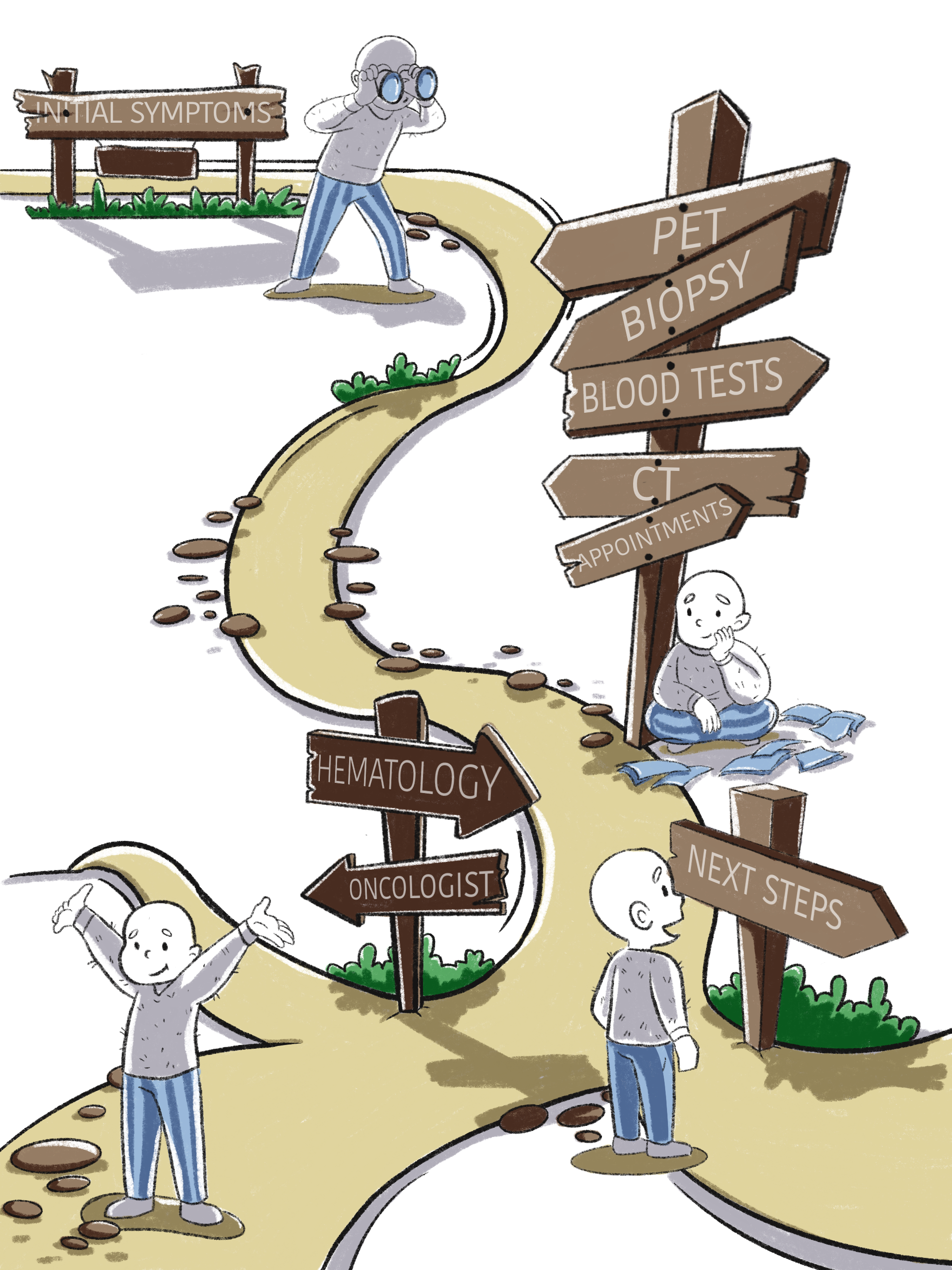
imaging might include:
X-ray
Ultrasound
CT Scan
MRI Scan
PET Scan
More information below

After imaging and blood tests you might need a biopsy
Taking a biopsy is the process of collecting a piece of tissue to look at the cells under the microscope and see if they are cancer cells.
There are different types of biopsies:
Needle or special device to take cells from the skin, usually with local numbing agents - Fine Needle Aspirate, Punch Biopsy
Some biopsies are done using imaging guidance to help the doctor find the right tissue (see imaging below for more information on imaging techniques) - Core Biopsy
Some biopsies require an operation to take a deeper or larger amount of skin tissue and might require a general anaesthetic (see surgery) - Open Biopsy
Based on the findings of the blood tests, imaging and biopsy, you might be referred to a specialist doctor for your type of cancer
Oncologist – specialises in solid organ cancers (eg lungs, kidney)
Haematologist – specialises in blood and lymph node cancers (eg leukaemia, lymphoma)
imaging
x-ray
X-ray beams are absorbed by the body tissues differently building a basic picture of internal bones and tissues

ultrasound
Uses sound waves emitted from a probe that are absorbed by tissues at different rates to build a picture of internal organs and tissues
ct scan
Takes many x-ray images combined together to form a 3D picture of internal organs and tissues
Much more detailed series of images compared to x-ray
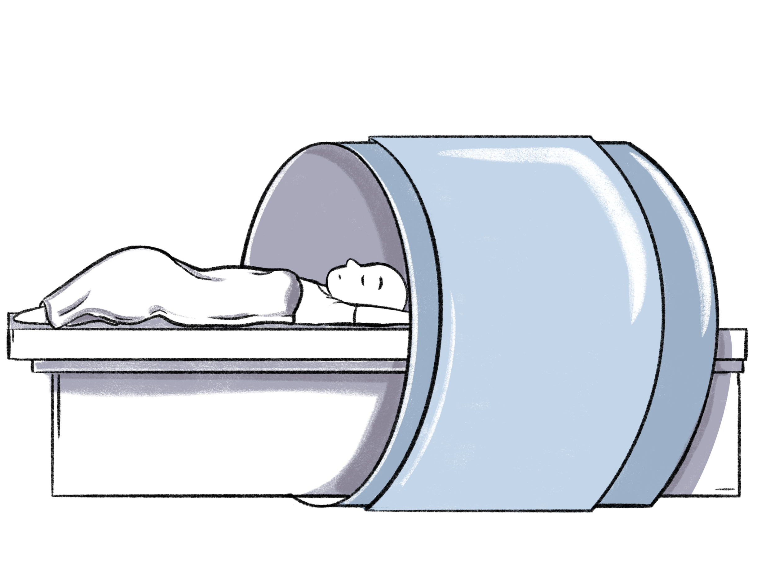
mri scan
Uses magnetic fields and radio waves through the area of interest to send a detailed 3D image of internal organs and tissues
Can be loud
Certain metals and implants inside the body (e.g pacemakers, cochlear implants) may not be able to go into the MRI scanner (ask your doctor)

pet scan
Uses a radioactive imaging dye (called a tracer) injected through a vein (usually in your arm) that is taken up by cells at a rate based on how active they are
More active cells can represent tumours or cancer cells